A Suspensory Way of Life: Integrating Locomotion, Postures, Limb Movements, and Forces in Two-Toed Sloths Choloepus didactylus
Granatosky, M. C., Karantanis, N. E., Rychlik, L., & Youlatos, D. (2018). A suspensory way of life: Integrating locomotion, postures, limb movements, and forces in two-toed sloths Choloepus didactylus (Megalonychidae, Folivora, Pilosa). Journal of Experimental Zoology Part A: Ecological and Integrative Physiology, 329(10), 570–588. https://doi.org/10.1002/jez.2221
Abstract
Over the last decade, we have learned much about the anatomy, evolutionary history, and biomechanics of the extant sloths. However, most of this study has involved studying sloths in controlled conditions, and few studies have explored how these animals are behaving in a naturalistic setting. In this study, we integrate positional activities in naturalistic conditions with kinematic and kinetic observations collected on a simulated runway to best capture the biomechanical behavior of Linnaeus’s two-toed sloths. We confirm that the dominant positional behaviors consist of hanging below the support using a combination of forelimbs and hindlimbs, and walking quadrupedally below the branches. The majority of these behaviors occur on horizontal substrates that are approximately 5–10 cm in diameter. The kinematics of suspensory walking observed both in the naturalistic settings and on simulated arboreal runways are dominated by movement of the proximal limb elements, while distal limb elements tend to show little excursion. Joint kinematics are similar between the naturalistic setting and the simulated runway, but movements of the shoulder and hip tend to be exaggerated while moving in simulated conditions. Kinetic patterns of the two-toed sloth can be explained almost entirely by considering them as an inverted linked strut. However, medially directed forces toward the substrate were more frequent than expected in the forelimb, which may help sloths maintain a better “grip” on the substrate. This study serves as a model of how to gain a comprehensive understanding of the functional-adaptive profile of a particular species.
Introduction
Among arboreal mammals, modern sloths (Choloepus spp. and Bradypus spp.) stand out by their remarkable, almost obligatory, commitment to a suspensory way of life. However, both genera seem to be well rooted within a group of basically fossorial, terrestrial, and semiarboreal extant and extinct relatives, rendering their specialized suspensory arboreality quite perplexing (Gaudin, 2004; Gaudin & McDonald, 2008; Pujos et al., 2017). In effect, despite their apparently similar behavioral adaptations, recent research has clarified that extant three-toed sloths (Bradypus spp.) and two-toed sloths (Choloepus spp.) are only distantly related, having separated between 23.5 and 39 mya (Ruiz-García et al., 2018; Slater et al., 2016), with the former considered to be either the sister group of all other extinct and extant sloths (Gaudin, 2004) or associated with extinct megatheriid sloths (Greenwood et al., 2001; Slater et al., 2016), and the latter belonging to the family Megalonychidae, consisting mainly of extinct ground sloths (McDonald & De Iuliis, 2008; Ruiz-García et al., 2018; Slater et al., 2016). It is thus parsimonious to consider that their morphofunctional and behavioral adaptations have evolved by convergence (see Nyakatura, 2012 for review).
Suspensory positional behavior extends the feeding sphere of arboreal animals, circumvents the danger of toppling over fragile, narrow, and flexible substrates, and thus provides access to a variety of microhabitats in forest tree canopies (Cartmill, 1985; Grand, 1972). However, two-toed and three-toed sloths, although sharing similar habitat attributes, seem to differentiate in their specific preferences: three-toed sloths are diurnal, prefer trees with large crowns, exposed to the sun and with moderate lianas, use vision-oriented exploration and appear to move at ease on generally larger and more vertical substrates, descend rump-first, and encircle large substrates with claws and volar pads, the latter contributing to friction for climbing (Mendel, 1985a; Montgomery & Sunquist, 1978). On the other hand, two-toed sloths are nocturnal, prefer larger trees with an abundant intertwined network of lianas, use olfaction-oriented exploration and mainly use small to medium-sized arboreal substrates, can descend head-first, and grasp large substrates with the tips of claws that enhance friction for climbing (Mendel, 1981a; Montgomery & Sunquist, 1978).
Despite these differences, both genera share some kinematic and morphological adaptations that relate to these specialized ways of life (Granatosky et al., 2014; Hodges et al., 2014; Mendel, 1979; Miller, 1935). Most of these observations derive mainly from studies on two-toed sloths conducted in laboratory conditions. During suspensory walking, interlimb coordination can be variable from lateral sequence/single foot gaits to diagonal sequence/single foot gaits at lower speeds, to trot-like diagonal couplets at higher speeds (Mendel, 1985a; Nyakatura et al., 2010). Speed of movement is initially increased by shortening both contact and swing duration, and subsequently by additionally extending step length (Nyakatura et al., 2010). Body progression is achieved primarily by retracting the most proximal elements of fore- and hindlimbs, which actually contribute mainly to step length (Nyakatura et al., 2010). Step length is further increased by bending movements of the thoracolumbar spine supported by large epaxial muscles (Gaudin & Nyakatura, 2017; Nyakatura & Fischer, 2010a).
Limb-loading patterns of the two-toed sloth are characterized by: (a) single-peak vertical forces that are evenly distributed between the forelimbs and hindlimbs (i.e., no forelimb or hindlimb-biased weight support as seen in most other mammals), (b) a propulsive force as the limb (forelimb or hindlimb) first touches down followed by a braking force before lift-off, (c) the forelimb tends to apply greater propulsive forces to the substrate, while the hindlimb tends to apply larger braking forces, and (d) high medially directed forces that approach, and in some cases exceed, the magnitude of the fore-aft forces (Granatosky & Schmitt, 2017). With the exception of the high medially directed forces, which may be a result of increased limb adduction and/or the bending of the spine, the limb-loading patterns of the two-toed sloth appear to reflect what is expected (with an appropriate minus sign) for a quadruped functioning as a linked strut with a center of mass (COM) at approximately 50% of trunk length (Bertram, 2016; Granatosky et al., 2018; Pontzer, 2007; Usherwood et al., 2007). This indicates that the kinetic patterns of sloths are passively determined by the position of the COM relative to the hands and feet (Bertram, 2016; Granatosky et al., 2018; Raichlen et al., 2009), and no active mechanisms (i.e., activation of extrinsic limb muscles to pitch the COM forward or backward via the horizontal lever effect [Larson & Demes, 2011; Larson & Stern, 2009; Reynolds, 1985a], or differential limb compliance [Schmitt, 1999; Schmitt & Hanna, 2004]) are used to modulate forces across the limbs (Young, 2012).
However, pilot data by Gorvet et al. (2018) demonstrates that three-toed sloths tend to activate the superficial pectoralis muscle, a forelimb protractor, during the early to middle portions of stance phase. Such behavior has the tendency to increase braking forces and shift weight closer to the forelimbs via the horizontal lever effect (see Reynolds, 1985a for discussion of this mechanism). Currently, no comprehensive data set has yet to determine whether two-toed sloths, or any suspensory quadruped, are using active or passive mechanisms to modulate limb-loading patterns during locomotion.
Suspensory movements and tensile loading of the limbs have been cited as having profound effects on the postcranial skeleton of living sloths. The limbs are elongated (Straus & Wislocki, 1932), with a rounded shape and low bone mineral density (Patel & Carlson, 2008; Patel et al., 2013). The relatively rigid hands and feet with large hooklike claws anchor flexed and perpendicularly on the branches (Mendel, 1981a, 1981b, 1985a), while the highly mobile wrist, the reduced olecranon, highly mobile pectoral girdle on the circular thorax, the ligamentous connection of the clavicle to the sternum, the highly mobile transverse tarsal joints, the low patellar trochlea, and the flexible hip joint promote extensive limb mobility and facilitate rotation, abduction and habitual flexion (Miller, 1935; Mendel, 1979, 1981a, 1981b, 1981c, 1985a; Nyakatura & Fischer, 2010b). These limb movements and postures are further powered by relatively strong shoulder and forelimb and hip and hindlimb flexors, with long tendinous distal insertions that increase their mechanical advantage (Fujiwara et al., 2011; Grand, 1978; Mendel, 1981a, 1981c, 1985a; Nyakatura & Fischer, 2010b).
Despite the amount of information on morphology and laboratory-based kinematics and kinetics, there are no quantitative observations on unrestrained locomotor and postural (=positional) behavior and associated limb postures of two-toed sloths in a naturalistic setting. Studies of positional behavior of either free-ranging animals or of captive animals within large enriched enclosures that simulate natural habitats inform on the frequency of occurrence and the performance of locomotor and postural modes under nonconstrained conditions and provide clues on their adaptive significance and morphological correlates. Positional data on free-ranging Bradypus variegatus in Venezuela demonstrated that three-toed sloths actually shared their locomotion between vertical climbing and under-branch suspensory locomotion, whereas above-branch sitting postures largely dominated over below-branch hanging (Urbani & Bosque, 2007). These findings partly support previous predictions and may actually account for the observed differences between three- and two-toed sloths (Mendel, 1981c, 1985b) that are translated in some morphological divergences (Grand, 1978; Miller, 1935; Nyakatura & Fischer, 2010a, 2010b; Olson et al., 2017). Therefore, comparable data on unrestrained locomotion and postures of two-toed sloths in a naturalistic setting would further contribute to the understanding of these morphological and behavioral differences and provide valuable information on the adaptive and evolutionary significance of suspensory arboreality.
In this context, this study provides the first quantitative analysis of locomotion, postures, and limb movements of captive Linnaeus’s two-toed sloths Choloepus didactylus, living in a large indoors enriched enclosure. These data are further combined with kinematic and kinetic data of suspensory walking collected on simulated arboreal supports to determine whether inferences drawn from laboratory-based findings are biologically valid. Furthermore, kinetic data are paired with kinematic patterns of limb movement to explore the extent that limb-loading patterns in two-toed sloths are determined by passive mechanisms, or potentially active modulation of forces. From this combination of data, we expect two-toed sloths (a) to be highly suspensory in both locomotor and postural terms, (b) to use small, medium, and horizontal substrates, (c) during locomotion to habitually use arm abduction and neutral-posture, elbow and wrist ulnar deviation, hip abduction and flexion, and knee flexion and foot abduction, (d) during postures to frequently maintain the arm in adduction and neutral-posture, the elbow in extension, the wrist in flexion, the hip in adduction and neutral-posture, the knee in extension, and the ankle abducted, (e) to use similar kinematic patterns during suspensory walking in a naturalistic environment and on a simulated arboreal support, and (f) to display kinetic patterns that are predictable by kinematics alone and not dependent on muscular activation for postural control for active weight-distribution between the limbs.
Materials and Methods
Locomotor and Postural Analysis
For the purposes of the current study, we observed and filmed one male and one female captive adult Linnaeus’s two-toed sloths Choloepus didactylus (Megalonychidae, Folivora, Pilosa) in the Nowe Zoo, Poznań, Poland. Both study animals are captive-born, healthy, fully habituated to human presence, and did not display any stereotypic behaviors during the study period. They are housed together in the display colonies of the Nocturnal Pavilion of the zoo. They inhabited a large enclosure (H: 300 cm × W: 500 cm × D: 300 cm) under a reversed day–night regime. The enclosure covers the innermost left side of the pavilion and is fronted by a large glass window. It contained a wide variety of available substrates of diverse sizes (diameters from 5 to 30 cm) and inclinations (horizontal to vertical), enabling the sloths to move freely in a three-dimensional enriched environment. The substrates involved branches, a fixed nest box, a hanging nest basket, and several hanging feeders that assured regular and ad libitum feeding of the study animals. Despite the enriched arboreal environment provided, substrates in an artificial enclosure are always expected to limit the positional options of caged animals. In this case, an estimate of their availability allows for a controlled test of substrate preference or avoidance. Therefore, we calculated all available substrates by unit and, subsequently, estimated the availability of the different size and inclination categories (see Table 1 for definitions). Small and medium substrates were rather equally represented (29.8% and 31.4%, respectively). Large and very large substrates were also equally represented (20.1% and 18.7%, respectively). Regarding substrate inclination categories, oblique substrates dominated (43.5%), whereas vertical and horizontal ones were more or less equally available (29.1% and 27.4%, respectively). Preference or avoidance of these categories was then estimated by Jacobs’ D value: D = (U − A)/(U + A − 2U × A), where U is proportion of use, and A is proportion of availability. Values of the index range from −1, depicting strong avoidance, to +1, showing strong preference, whereas values around 0 are considered as neutral.
The present data derive from the analysis of extensive video recordings of the study animals. For the experimental procedure, the Nowe Zoo administration granted a research permit to film the animals in situ between February and May 2013, during the days when the zoo was closed to the general public. This allowed easy and uninterrupted access to the animal enclosure without external disturbance. For these administrative reasons, the animals were filmed twice per week from 10:00 to 17:00, using additional infrared lighting. During video recording sessions we used a SONY Hi-8 CCD-TR705E camcorder, at 24 fps and at a shutter speed of 1/500th. The original Hi-8 tapes, totaling 32 hr of recordings, were digitized and were subsequently analyzed frame-by-frame on a PC for data collection.
During video analyses, we used a 30-s scan sampling for data collection from all visible individuals (Martin & Bateson, 1993). This time lapse was estimated as sufficient for relatively independent events of locomotor and postural behavior for such slow-moving animals. During each scan instant we recorded: (a) behavioral context, (b) substrate size, (c) substrate inclination, (d) locomotor or postural mode, (e) arm adduction/abduction, (f) arm protraction/retraction, (g) elbow flexion/extension, (h) wrist radial/ulnar deviation, (i) hip adduction/abduction, (j) hip protraction/retraction, (k) knee flexion/extension, and (l) ankle abduction/adduction (see Table 1 for the different categories of the recorded variables and Figure 1 for the most common modes). At the wrist and ankle joints we opted for radial/ulnar deviation and abduction/adduction, respectively, as sloths grasp with inverted hand and foot postures (Mendel, 1981c). Substrate size categories were defined in respect to the size of the hands and feet of the animals. Furthermore, although positional modes and substrate use usually relate to behavioral contexts (e.g., feeding, traveling, etc.) in the wild, a captive setting with its spatial limitations and specific feeding conditions usually modifies and biases similar associations.
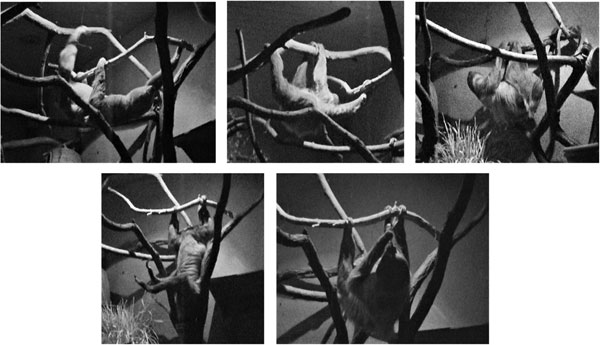
Table 1. Definition and description of recorded variables for captive Choloepus didactylus.
| Category | Variant | Description |
|---|---|---|
| Substrate size | Small | Diameter ≤ 5 cm |
| Medium | 5 cm < diameter ≤ 10 cm | |
| Large | 10 cm < diameter ≤ 20 cm | |
| Very large | Diameter > 20 cm | |
| Substrate inclination | Horizontal | 0°–22.5° |
| Oblique | 22.5°–67.5° | |
| Vertical | 67.5°–90° | |
| Locomotor mode | Suspensory walk | Below branch quadrupedal suspension involving regular movements of the limbs along a single substrate |
| Suspensory clamber | Below branch quadrupedal suspension involving irregular movements of the limbs along multiple substrates | |
| Bridge | Below branch suspensory reaching across distantly located substrates involving irregular limb movements | |
| Vertical ascent/descent | Upwards (head-first) and downwards (head- or rump-first) vertical displacement along a single substrate involving regular limb movements | |
| Pronograde walk/clamber | Above branch quadrupedal displacement involving regular or irregular limb movements along a single, or across multiple substrates | |
| Postural mode | Forelimb and hindlimb suspension | Below branch suspensory postures involving combination of fore- and hindlimbs |
| Hindlimb-only suspension | Below branch suspensory postures involving only one or both hindlimbs | |
| Forelimb-only suspension | Below branch suspensory postures involving only one or both forelimbs | |
| Sit | Above branch seated posture | |
| Lie | Above branch reclining with limbs hanging freely | |
| Cling | Clinging with flexed fore- and hindlimbs along a single vertical or steeply inclined substrate | |
| Arm abduction/adduction | Abduction | Arm kept away from the sagittal plane |
| Adduction | Arm kept close to the sagittal plane | |
| Hip abduction/adduction | Abduction | Thigh kept away from the sagittal plane |
| Adduction | Thigh kept close to the sagittal plane | |
| Arm protraction/retraction | Protraction | Arm extended above the shoulder |
| Neutral-posture | Arm kept at the level of the shoulder | |
| Retraction | Arm flexed below the shoulder | |
| Hip protraction/retraction | Protraction | Thigh flexed |
| Neutral-posture | Thigh kept at the level of the hip | |
| Retraction | Thigh extended | |
| Elbow flexion/extension | Flexion | Elbow flexed |
| Extension | Elbow extended | |
| Knee flexion/extension | Flexion | Knee flexed |
| Extension | Knee extended | |
| Wrist radial/ulnar deviation | Radial deviation | Wrist turned radially |
| Ulnar deviation | Wrist turned ulnarly | |
| Ankle abduction/adduction | Adduction | Upper ankle joint adducted |
| Abduction | Upper ankle joint abducted |
At the end of the sampling process, the total of collected instants of locomotor and postural behavior was derived from the observational recording of the two different individual sloths. We preferred to combine the observational data, as sometimes we were not certain of the identity of the sampled animals. Furthermore, a common problem of positional behavior studies is the autocorrelation of successive sampling events. This occurs because subsequent samples of the same individuals usually lack independence from previous observations (Dawkins, 2007). To address this shortcoming and safely guarantee independence, we followed a trimming procedure. Initially, the complete data set was divided into locomotor and postural subsets. Then, in each subset, we only considered every second instant (i, i + 2), deleting each intermediate one (i + 1). Following this trimming procedure, we obtained a total of 948 counts of locomotion and 956 counts of postures. Differences among frequencies of behaviors or substrate use were calculated using log-likelihood G tests. P-values of 0.05 or less were regarded as statistically significant and only those are reported in the results section. The present research followed the guidelines for the treatment of animals in behavioral research and teaching (ASAB/ABS, 2012) and complied with the regulations and legislations of the Nowe Zoo, and the Adam Mickiewicz University in Poznań, and the legislation of the Aristotle University of Thessaloniki.
Kinetic and Kinematic Analysis
Kinematic and kinetic data were collected from two different subjects of two-toed sloths (Choloepus didactylus) at the Central Florida Zoo (Sanford, FL) following the protocols approved by Duke University Institutional Animal Care and Use Committee (IACUC protocol #A270-11-10). Both animals were adults and were clear of any pathologies or gait abnormalities (Table 2).
Table 2. Descriptive summary and number of steps analyzed (where applicable) of the studied subjects of Choloepus didactylus.
| Location | Subject | Sex | Body mass (kg) | Date of birth | Forelimb steps analyzed | Hindlimb steps analyzed |
|---|---|---|---|---|---|---|
| Central Florida Zoo (United States) | Individual 1 | Female | 7.25 | 11/09/2000 | 9 | 9 |
| Central Florida Zoo (United States) | Individual 2 | Female | 6.85 | 05/08/2003 | 16 | 9 |
| Nowe Zoo Poznań (Poland) | Individual 1 | Male | 7.10 | 30/07/1995 | – | – |
| Nowe Zoo Poznań (Poland) | Individual 2 | Female | 6.95 | 06/03/2004 | – | – |
Data were collected while animals walked below an instrumented runway measuring 366 cm in length and 3.10 cm in diameter. During all trials, animals were video-recorded from a lateral view at 120 fields/s using a GoPro camera (Hero 3+ Black Edition; GoPro, San Mateo, CA) modified with a Back-Bone Ribcage (Ribcage v. 1.0; Back-Bone, Ottawa, ON, Canada), which allows the GoPro cameras to be outfitted with interchangeable lenses and eliminates image distortion inherent to the camera (Granatosky et al., 2016). Only walking strides (i.e., duty factor over 50%) in which the animal was traveling in a straight path and not accelerating or decelerating (i.e., steady-state locomotion) were selected for analysis. Steady-state locomotion was determined by calculating the instantaneous velocity between subsequent video frames throughout the entire stride, and then using regression analysis to determine whether velocity changed throughout the stride (Granatosky, 2015; Granatosky & Schmitt, 2017; Granatosky et al., 2016). Only strides with no change in speed (i.e., slope not significantly different from zero) were analyzed. All statistical tests described below were conducted in MATLAB (MathWorks, Natick, MA).
From video recordings, the position of the shoulder, elbow, wrist, hip, knee, ankle, and forelimb and hindlimb point-of-contact (POC) were collected over the course of support phase. The resulting x–y coordinate data were used to track angular movements in the shoulder, elbow, wrist, hip, knee, and ankle. All limb angles were digitized using DLT Dataviewer (Hedrick, 2008) in MATLAB.
All angular movements were measured in degrees (°). To make joint movements comparable between strides and different individuals, all joint data were scaled as a percentage of support phase. Following Larson et al. (2000) shoulder and hip angles were measured relative to the vertical axis of the shoulder or hip joint (i.e., when the arm passed directly above or below [depending on orientation] the shoulder or hip joint this was considered the neutral position [0°]). Angles greater than 15° represent shoulder or hip protraction, while angles less than −15° represent shoulder or hip retraction. Angles between −15° and 15° represent shoulder or hip neutral-posture. Elbow and knee angles were measured so that 180° represents maximum elbow or knee extension. Angles greater than 90° represent elbow or knee extension, while angles less than 90° represent elbow or knee flexion. For movements of the wrist and ankle, angles were measured based on the position of the wrist relative to the POC with the support and the elbow. Neutral position (180°) was defined as the point in which the wrist or ankle was in line with the POC and the elbow or knee. Because sloths walk with inverted hand and foot positions, traditional terms like flexion/extension and dorsiflexion/plantarflexion do not accurately describe kinematic patterns of movement. For the wrist, angles greater than 225° represent radial deviation, while angles less than 225° are ulnar deviation. For the ankle, angles greater than 225° represent adduction, while angles less than 225° represent abduction. Because only lateral view cameras were used to collect kinematic data it was not possible to collect angular data for arm and hip abduction and adduction. Accordingly, arm and hip positions were scored as abducted or adducted at 10% intervals during stance phase. From these data, we determined the frequency of arm and hip abduction and adduction during suspensory walking on the simulated arboreal runway.
To assess whether sloths use similar kinematic patterns during suspensory walking in a naturalistic setting and on a simulated arboreal support, we quantified angular data of joint movements collected during stance phase into frequencies of: (a) arm adduction/abduction, (b) arm protraction/retraction, (c) elbow flexion/extension, (d) wrist radial/ulnar deviation, (e) hip adduction/abduction, (f) hip protraction/retraction, (g) knee flexion/extension, and (h) ankle adduction/abduction. To make comparisons as biologically relevant as possible, we compared kinematic frequencies on the simulated arboreal runway only to data collected on horizontally positioned small and medium diameter substrates in the naturalistic enclosure. We used a χ2 goodness of fit test to determine whether the frequency of joint positions used by two-toed sloths during suspensory walking on the simulated arboreal support differed significantly from what was observed during suspensory walking in the naturalistic setting.
To collect kinetic data, a small subsection of the runway was instrumented with two Kistler force plates (model 9317B; Kistler, Amherst, NY) that have been used in previous studies (Granatosky & Schmitt, 2017; Granatosky et al., 2016). Force plate output was sampled at 12,000 Hz, imported, summed, and processed using BioWare™ v.5.1 software, and then filtered (second-order Low Pass Butterworth, 30 Hz) in MATLAB. Only steps with single-limb contacts on the plate or those steps in which the forelimb and hindlimb forces could be clearly differentiated were analyzed. From force plate data, vertical impulse (VI), the time at which the propulsive to braking transition (PB) occurred, and whether the limb was exerting medially or laterally directed force were determined for each limb single limb contact. Vertical impulse was measured as the area under the force–time curve in the vertical component of the substrate reaction force. To make statistical comparisons between subjects of differing body masses, all impulses were measured as a percentage of body weight seconds (%bws). The PB was defined as the point that the fore-aft force switches from a propulsive (positive) to braking (negative) force. Medially and laterally directed forces were measured from the mediolateral force trace. Medially directed forces were defined as those where the animal was applying a negative force on to the substrate, while laterally directed forces were the opposite.
To determine whether two-toed sloths are using passive or active mechanisms to modulate force patterns we used three separate analyses that integrate kinetic and kinematic data. All of these analyses assume that passive mechanisms are being used to modulate forces, so deviations from these expected patterns would indicate that sloths are using active mechanisms to modulate forces.
Our first test to assess whether two-toed sloths are using passive or active mechanisms to modulate force patterns has been assessed by Pontzer (2007), Usherwood et al. (2007), Bertram (2016), and Granatosky et al. (2018), and tests the relationship between PB and COM position. Specifically, these authors suggest that quadrupeds are best modeled passively as a set of linked struts with the COM lying somewhere in the middle (Bertram, 2016; Usherwood et al., 2007). In a linked strut model, animals apply forces that act along a line from the POC to the COM (Bertram, 2016). Thus, the magnitude of the horizontal force will be equal to the vertical force multiplied by the tangent of the angle between a line from the POC to the COM and the vertical. In this model, the PB will occur at the time that the COM passes the POC. Our ability to test whether sloths behave as linked struts was constrained by the availability of previously published data on COM position in two-toed sloths. We calculated COM position for two-toed sloths using the segment mass method described by Preuschoft and Demes (1984) and Vilensky (1978) based on segment parameters provided by Nyakatura and Andrada (2013) and static images (N = 25) of sloths locomoting from our video-recordings. From these calculations the COM position for a two-toed sloth is approximately 50.4 ± 1.6% of trunk length, as measured from the base of the skull.
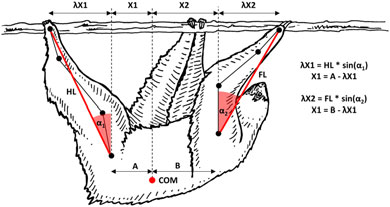
After COM position was determined, we digitized the position of the COM along the trunk, and for each single limb contact collected the timing of touchdown, the point at which the COM passed beneath the POC, and lift-off. We then calculated the proportion of support phase at which the COM passed beneath the POC for each single limb contact. It should be noted that these COM positions were collected from animals not used in this study and that the COM does not remain static during locomotion (but see Farrell et al., 2015 and Young et al., 2007 who suggest anterior/posterior movement appears to be minimal). Taken together, it is possible that potential errors in the timing of events may have occurred. We conducted a paired t test on ranked values to determine the statistical likelihood of the PB and the point at which the COM passed beneath the POC occurring at the same time for each limb separately.
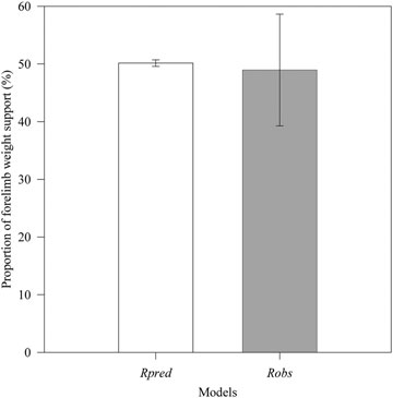
The second test to determine whether two-toed sloths are using passive or active mechanisms to modulate force patterns between the limbs was to explore the potential that changes in limb position relative to the COM will influence vertical force distribution between the limbs (Gray, 1944, 1968; Larson & Demes, 2011; Raichlen et al., 2009; Reynolds, 1985a; Young, 2012). If sloths are using passive mechanisms to modulate forces, the limb that is closest to the COM will support a greater proportion of body weight (Larson & Demes, 2011; Raichlen et al., 2009; Young, 2012). To test this, we followed methods used by Raichlen et al. (2009) and Larson and Demes (2011) in which the observed vertical force distribution (i.e., the vertical impulse ratio (Robs)) was compared to the predicted distribution of vertical force (Rpred) based on the average position of the forelimb and hindlimb POC relative to the COM (Figure 2). We calculated Robs as VIFL / VItotal, where VIFL is the VI in the forelimb, and VItotal is the sum of the VI in the forelimb and hindlimb within the same stride. Values equal to 0.5 represent equal weight distribution between the limbs. Values lower than 0.5 indicate greater weight distribution borne by the hindlimbs, and values greater than 0.5 indicate greater weight distribution borne by the forelimbs. Rpred was calculated as X1 / (X1 + X2), where X1 is the average horizontal distance from the hindlimb POC to the COM during hindlimb support and X2 is the average horizontal distance from the forelimb POC to the COM during forelimb support, as calculated from the video-recordings. We conducted a paired t test on ranked values to determine the statistical likelihood of Robs and Rpred being the same. It should be noted that the ability to conduct this analysis depends upon the ability to collect both forelimb and hindlimb forces within a single stride. This limited our analysis to only a subset of the originally collected kinematic data.
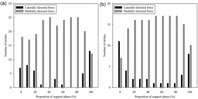
Finally, we compared the association between mediolateral forces and whether the limb was in an adducted or abducted position. Mediolateral force traces were scored as either being medially directed (a negative value on the mediolateral force curve) or laterally directed (a positive value on the mediolateral force curve) at 10% intervals during stance phase. We used χ2 goodness of fit tests to determine whether the frequency of medially and laterally directed forces differed significantly from when the limb was in an adducted or abducted position.
Results
Positional Profile
During the study period, sloths moved freely within the enclosure and used many different arboreal pathways as provided by the availability and the setting of the different types of substrates. Thus, in terms of substrate size, captive sloths mainly used and preferred medium substrates (D = 0.49; Figure 3). Large substrates were also considerably used, and their use increased significantly during locomotion (G = 6.908, p = 0.035). Small and very large substrates were used less and were moderately and strongly avoided, respectively (D = −0.27 and D = −0.91, respectively). Overall, horizontal substrates were largely used (Figure 3) and strongly preferred (D = 0.68). Oblique substrates were also adequately used but were lightly avoided (D = −0.27). On the other hand, vertical substrates were seldom used and were strongly avoided (D = −0.89). Postural behavior was marked by a significant increase of horizontal substrate use (G = 32.5, p < 0.0001). In contrast, during locomotion, the use of oblique substrates showed significant increase (G = 13.8, p = 0.0002).
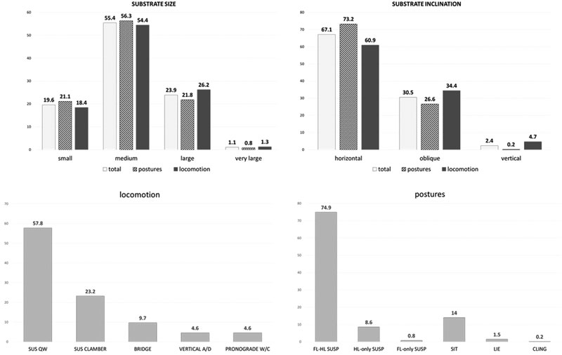
Below branch suspension were the dominant postures and represented 84.3% of all postural counts (Figure 3). In contrast, above branch postures (lie and sit) scored quite low (15.5%). Dominant suspensory postures were those involving at least one combination of forelimb and hindlimb (88.9% of suspensory postures subsample). Hindlimb-only hanging was also used (10.2% of suspensory postures subsample), but forelimb-only hanging was rare (0.9% of suspensory postures subsample). Suspensory quadrupedal walking was the most frequent locomotor mode, followed by suspensory quadrupedal clamber (Figure 3). Bridging between gaps of substrates was also regular and vertical ascents and descents were seldom used. Pronograde quadrupedal above branch locomotion was also observed, but at low rates (Figure 3).
Limb Postures
Overall, the arm was mainly adducted and placed at neutral-posture relative to the shoulder joint (Table 3). However, adduction was significantly more frequent during retraction of the arm (G = 20.6, p < 0.0001; Table 4). In general, the elbow was equally flexed and extended (Table 3), and this was also mainly the case, when the arm was in neutral-posture (Table 4). In contrast, when the arm was protracted the elbow was more frequently extended (G = 45.5, p < 0.0001) and when the arm was retracted, it was habitually flexed (G = 30.7, p < 0.0001; Table 4). Finally, the wrist was basically ulnarly deviated (Table 3). The wrist was also kept ulnarly deviated when the elbow was extended, but there was a slight increase in wrist radial deviation when the elbow was flexed (G = 19.8, p < 0.0001; Table 4).
Regarding the hindlimb, the hip was mostly abducted and habitually flexed (Table 5). Abduction was particularly frequent during hip extension (G = 27.6, p < 0.0001) and to a lesser extent, during hip flexion (G = 15.9, p = 0.0001; Table 4). On the other hand, adduction was dominant in hip neutral-posture (G = 23.6, p < 0.0001; Table 4). In general, the knee was regularly flexed (Table 5). Knee flexion was especially common during hip flexion (G = 81.8, p < 0.0001), whereas knee extension was more frequent in hip extension (G = 50.5, p < 0.0001) and a hip neutral-posture (G = 5.7, p = 0.003; Table 4). Overall, the ankle was mainly kept abducted (Table 5). This posture was very frequent when the knee was extended, but there was a slight, but significant, increase in ankle adduction during knee flexion (G = 47.3, p < 0.0001; Table 4).
Table 3. Percentages of forelimb postures during postural behavior, locomotion, and the major positional modes in captive Choloepus didactylus (total sample corresponds to all counts where the forelimb was in contact with a substrate).
| Posture | Total | Postures | Locomotion | Susp. postures | Susp. clamber | Bridging | Susp. walk |
|---|---|---|---|---|---|---|---|
| Arm abduction | 39.4 | 33.2 | 45.2 | 31.2 | 69.1 | 65.2 | 26.3 |
| Arm adduction | 60.6 | 66.8 | 54.8 | 68.8 | 30.9 | 34.8 | 73.7 |
| Arm protraction | 26.1 | 20.4 | 31.4 | 19.1 | 29.1 | 47.8 | 31.7 |
| Arm neutral-posture | 42.4 | 46.2 | 38.8 | 45.9 | 40.0 | 34.8 | 39.8 |
| Arm retraction | 31.5 | 33.4 | 29.8 | 35.1 | 30.9 | 17.4 | 28.5 |
| Elbow flexion | 50.4 | 42.8 | 57.4 | 33.2 | 72.7 | 52.2 | 47.4 |
| Elbow extension | 49.6 | 57.2 | 42.6 | 66.8 | 27.3 | 47.8 | 52.6 |
| Wrist ulnar deviation | 95.5 | 97.5 | 93.7 | 100.0 | 98.2 | 93.5 | 99.6 |
| Wrist radial deviation | 4.5 | 2.5 | 6.3 | 0.0 | 1.8 | 6.5 | 0.4 |
| n | 1,822 | 874 | 948 | 724 | 220 | 92 | 548 |
Forelimb postures displayed some significant differences between locomotion and postures. In general, the arm was significantly more frequently adducted during postures than during locomotion (G = 27.4, p < 0.0001; Table 3). Increased arm adduction rates were also observed during suspensory postures and suspensory walk, whereas arm abduction dominated during suspensory clamber and bridging (G = 162.0, p < 0.0001; Table 3). An arm neutral-posture was frequent during both locomotion and postural behavior (Table 3). However, its percentage significantly decreased (G = 10.2, p = 0.0015), and arm protraction significantly increased during locomotion (G = 29.1, p < 0.0001; Table 3). An arm neutral-posture was the dominant limb posture during suspensory postures, suspensory walk and suspensory clamber, whereas bridging was characterized by significantly frequent arm protraction (G = 51.3, p < 0.0001; Table 3). Arm protraction was significantly more frequent in both suspensory clamber and walk compared to suspensory postures (clamber vs. postures: G = 9.6, p = 0.0082; walk vs. postures: G = 27.2, p < 0.0001; Table 3).
Elbow flexion was more common during locomotion, but elbow extension scored significantly higher during postures (G = 38.9, p < 0.0001; Table 3). Elbow flexion was very frequent during suspensory postures and quite frequent during suspensory walk, but elbow flexion was quite common during bridging and very common during suspensory clamber (G = 114.8, p < 0.0001; postures vs. walk: G = 26.7, p < 0.0001; clamber vs. bridging: G = 12.0, p = 0.0006; Table 3).
In both postures and locomotion, the wrist was maintained ulnarly deviated, but rates of radial deviation increased significantly during locomotion (G = 16.0, p = 0.0001; Table 3). Ulnar deviation of the wrist was also dominant during suspensory postures, suspensory walk, suspensory clamber, and bridging (Table 3).
Table 4. Fore- and hindlimb posture combinations during positional behavior of captive Choloepus didactylus (all data reported as a percentage).
| Arm protraction | Arm neutral-posture | Arm retraction | Hip protraction | Hip neutral-posture | Hip retraction | ||
|---|---|---|---|---|---|---|---|
| Arm abduction | 40.3 | 46.4 | 29.3 | Hip abduction | 52.5 | 39.3 | 69.1 |
| Arm adduction | 59.7 | 53.6 | 70.7 | Hip adduction | 47.5 | 60.7 | 30.9 |
| n | 238 | 386 | 287 | 505 | 257 | 188 | |
| Elbow flexion | 23.9 | 50.8 | 71.8 | Knee flexion | 70.1 | 45.1 | 35.6 |
| Elbow extension | 76.1 | 49.2 | 28.2 | Knee extension | 29.9 | 54.9 | 64.4 |
| n | 238 | 386 | 287 | 188 | 257 | 505 |
| Elbow flexion | Elbow extension | Knee flexion | Knee extension | |||
|---|---|---|---|---|---|---|
| Wrist ulnar deviation | 92.6 | 98.5 | Ankle abduction | 91.2 | 99.8 | |
| Wrist radial deviation | 7.4 | 1.5 | Ankle adduction | 8.8 | 0.2 | |
| n | 459 | 452 | 537 | 413 |
Limb Postures During Locomotion and Postures
In a similar manner to the forelimb, hindlimb postures differed between locomotor and postural behavior. The hip was mainly adducted during postures but was significantly kept more frequently abducted during locomotion (G = 40.4, p < 0.0001; Table 5). Hip adduction and abduction were shared in suspensory postures, and adduction was slightly more frequent in suspensory walk (G = 3.6, p = 0.0333; Table 5). In contrast, hip abduction was dominant during suspensory clamber and bridging (postures vs. clamber: G = 44.3, p < 0.0001; postures vs. bridging: G = 61.4, p < 0.0001; Table 5).
Although hip protraction was dominant during both locomotion and postures, hip retraction was more frequent during locomotion (G = 26.3, p < 0.0001), and a hip neutral-posture was more frequent during postural behavior (G = 41.9, p < 0.0001; Table 5). Hip protraction and neutral-posture were frequently observed during suspensory postures, but hip protraction was the dominant posture during suspensory walking and clamber (postures vs. walk: G = 17.3, p = 0.0002; postures vs. clamber: G = 7.3, p = 0.0291; Table 5). On the other hand, hip protraction and retraction were frequently used during bridging (bridge vs. postures: G = 35.9, p < 0.0001; bridge vs. walk: G = 18.7, p = 0.0002; bridge vs. clamber: G = 14.4, p = 0.0013; Table 5).
Knee flexion was largely dominant during postures but rates of flexion and extension were more equally shared during locomotion (G = 15.0, p = 0.0001; Table 5). Shared rates of flexion and extension were observed during suspensory postures and bridging (Table 4). On the other hand, knee extension was dominant during suspensory walk, and knee flexion was very frequent during suspensory clamber (G = 83.9, p < 0.0001; Table 5).
In the ankle joint, there were no differences between locomotion and postures, with the foot primarily kept in abduction (Table 5). Abduction is by far the dominant foot posture during suspensory postures, suspensory walk, suspensory clamber, and bridging (Table 5).
Table 5. Percentages of hindlimb postures during postural behavior, locomotion, and the major positional modes in captive Choloepus didactylus (total sample corresponds to all counts where the hindlimb was in contact with a substrate).
| Posture | Total | Postures | Locomotion | Susp. postures | Susp. clamber | Bridging | Susp. walk |
|---|---|---|---|---|---|---|---|
| Hip abduction | 52.2 | 45.0 | 59.5 | 48.9 | 73.6 | 89.1 | 43.8 |
| Hip adduction | 47.8 | 55.0 | 40.5 | 51.1 | 26.4 | 10.8 | 56.2 |
| Hip protraction | 53.2 | 51.2 | 55.1 | 49.1 | 52.7 | 47.8 | 55.8 |
| Hip neutral-posture | 27.0 | 33.6 | 20.4 | 33.3 | 24.6 | 10.9 | 23.0 |
| Hip retraction | 19.8 | 15.1 | 24.5 | 17.5 | 22.7 | 41.3 | 21.2 |
| Knee flexion | 56.5 | 60.9 | 52.1 | 53.4 | 73.6 | 54.3 | 37.6 |
| Knee extension | 43.5 | 39.1 | 47.9 | 46.6 | 26.4 | 45.7 | 62.4 |
| Ankle abduction | 94.9 | 94.5 | 95.4 | 100.0 | 99.1 | 100.0 | 99.3 |
| Ankle adduction | 5.1 | 5.5 | 4.6 | 0.0 | 0.9 | 0.0 | 0.7 |
| n | 1,900 | 952 | 948 | 798 | 220 | 92 | 548 |
Kinematic Comparisons Between a Naturalistic Setting and Simulated Arboreal Substrate
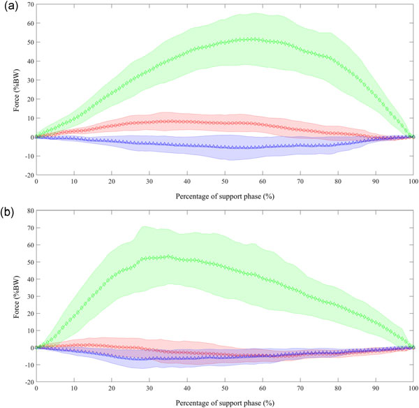
During suspensory walking on the simulated arboreal runway, the arm tended to be in an abducted position during the early portions of support phase. As progression continued, the arm was brought close to the body in an adducted position and remained that way until the end of support phase (Figure 4). At the same time, the arm was placed in a neutral-posture position at touchdown, and as support phase continued the arm was retracted substantially and approached the horizontal axis of the body (i.e., shoulder angle at 90°; Figure 5). Elbow positions at touchdown began in a relatively extended position and subsequently flexed until approximately 70% of support phase. The elbow then began to re-extend until the end of support phase. True elbow flexion, as defined by our criteria, was only observed approximately 35% of support phase (Table 3). The wrist was always kept in an ulnarly deviated position during suspensory walking on the simulated arboreal runway, and joint excursion was minimal. Joint positions during suspensory quadrupedal walking on the simulated arboreal runway and in the naturalistic enclosure proved to be generally similar for arm adduction/abduction and for wrist ulnar/radial deviation. Significant differences were observed in the frequency of arm protraction/retraction (df = 2, χ2 = 58.6, p ≤ 0.001) and elbow flexion/extension between the two conditions (df = 1, χ2 = 7.95, p ≤ 0.001). These differences stem from the generally more retracted arm and extended elbow positions observed while walking on the simulated arboreal support (Table 6).
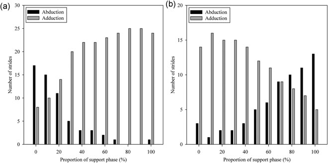
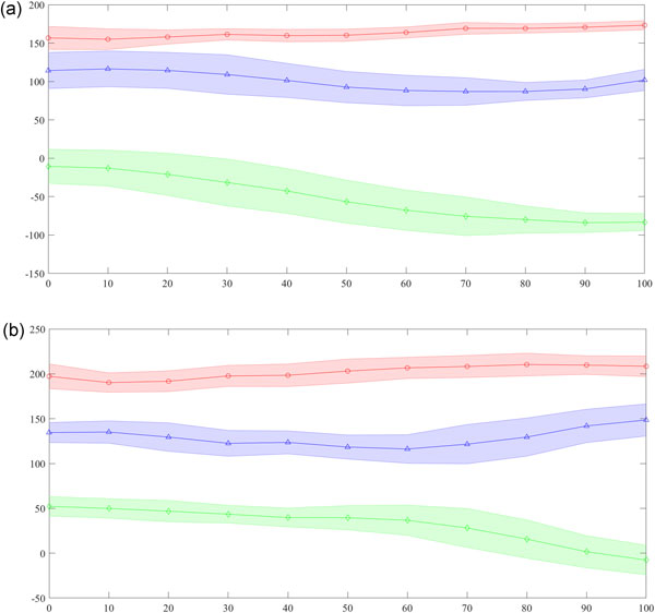
Table 6. Percentages of forelimb and hindlimb postures in captive Choloepus didactylus during suspensory walking on horizontally positioned small and medium diameter substrates in the naturalistic enclosure and on the simulated arboreal runway.
| Forelimb kinematics | Susp. walk | Susp. walk (runway) | Hindlimb kinematics | Susp. walk | Susp. walk (runway) |
|---|---|---|---|---|---|
| Arm abduction | 32.84 | 21.1 | Hip abduction | 49.25 | 32.8 |
| Arm adduction | 67.16 | 78.9 | Hip adduction | 50.75 | 67.2 |
| Arm protraction | 30.60 | 5.2 | Hip protraction | 52.99 | 77.49 |
| Arm neutral-posture | 43.28 | 15.2 | Hip neutral-posture | 26.12 | 16.45 |
| Arm retraction | 26.12 | 79.7 | Hip retraction | 20.90 | 6.06 |
| Elbow flexion | 54.48 | 35.1 | Knee flexion | 40.30 | 0.40 |
| Elbow extension | 45.52 | 64.9 | Knee extension | 59.70 | 99.6 |
| Wrist ulnar deviation | 99.25 | 100 | Ankle abduction | 99.25 | 94.8 |
| Wrist radial deviation | 0.75 | 0 | Ankle adduction | 0.75 | 5.2 |
| n | 268 | 25 | n | 268 | 18 |
In contrast to the patterns observed in the forelimb, the hip tended to be in an adducted and protracted position during most of support phase and was swung out in an abducted and neutral-posture/retracted position only near the end of support phase (~80%) while animals walked on the simulated arboreal substrate (Figures 4 and 5). Similar to the elbow, the knee was kept in a relatively extended position throughout support phase. Animals tended to flex the knee slightly near midsupport, but then re-extended it before lift-off. The ankle moved very little and was almost always kept in an abducted position while animals walked on the simulated arboreal support (Figure 5). Joint positions during suspensory quadrupedal walking on the simulated arboreal runway and in the naturalistic enclosure proved to be generally similar for ankle adduction/abduction. Significant differences were observed in the frequency of hip adduction/abduction (df = 1, χ2 = 5.59, p = 0.018) and protraction/retraction (df = 2, χ2 = 14.97, p ≤ 0.001) and knee flexion/extension (df = 1, χ2 = 49.01, p ≤ 0.001) between the two conditions. These differences stem from the generally more adducted and protracted hip and extended knee positions observed while walking on the simulated arboreal support (Table 6).
Testing Models of Passive Force Modulation
Statistical analysis of the association between the occurrence of the PB and the point at which the estimated COM passed under the forelimb/hindlimb POC revealed that there was no statistically significant difference in the occurrence of these events in either the forelimb or the hindlimb for two-toed sloths (Table 7). Furthermore, comparisons between Rpred (50.2 ± 0.6, N = 13) and Robs (50.0 ± 9.7, N = 13) revealed no significant difference (Figures 6 and 7). Finally, when comparing the relative frequencies between whether a limb was adducted/abducted and applying medially/laterally directed force the only significant (df = 10, χ2 = 17.5, p = 0.04) difference observed was between the frequency of the arm in an abducted position and the frequency of the arm applying a laterally directed force. This finding indicates that in many instances throughout support phase a medially directed force was being applied to the substrate although the arm was in an abducted position (Figure 8).
Table 7. The timing (mean ± standard deviation; in s) and p value of the propulsive to braking transition and the portion of support phase where COM passes limb POC for captive Choloepus didactylus.
| Limb | N | PB | Portion of support phase where COM passes limb POC | Comparison p value |
|---|---|---|---|---|
| Forelimb | 25 | 82.7 ± 10.8 | 87.5 ± 8.4 | 0.151 |
| Hindlimb | 18 | 21.0 ± 14.9 | 17.9 ± 7.6 | 0.987 |
Discussion
Over the last decade, a wealth of information has been produced about the anatomy, evolutionary history, and biomechanics of the extant tree sloths (Billet et al., 2012; Cliffe et al., 2014; Fujiwara et al., 2011; Gaudin, 2004; Gorvet et al., 2018; Granatosky & Schmitt, 2017; Hodges et al., 2014; Nyakatura, 2012; Nyakatura & Andrada, 2013; Nyakatura & Fischer, 2010b; Nyakatura et al., 2010). From these data, we have greatly increased our knowledge about these animals and challenges of living a life upside-down (Granatosky & Schmitt, 2017; Nyakatura, 2012; Nyakatura et al., 2010) and in slow motion (Usherwood & Self Davies, 2017). However, until this study no one since Mendel (1981a, 1981c, 1985b) has stepped back to see how these animals are behaving in a naturalistic setting, and whether habitat-use and anatomical configuration influence the biomechanical performance of these animals. In this study, the two sets of data derived from different animals (naturalistic setting: sloths in Nowe Zoo Poznań; kinematic and kinetic experiments: sloths in Central Florida Zoo). Nevertheless, we are confident that our results reflect the behavioral and biomechanical profiles of the species, and the comparable methods used in the two settings enable their combination into an integrated view of the arboreal suspensory behavior of two-toed sloths.
In this study, we confirm, rather unsurprisingly, that the dominant postural behaviors of two-toed sloths consist of hanging below the support using a combination of forelimbs and hindlimbs and walking quadrupedally below the branches. The majority of these behaviors occur on horizontal substrates that are approximately 5–10 cm in diameter. The kinematics of suspensory walking observed both in the naturalistic settings and on simulated arboreal runways are dominated by movement of the proximal limb elements, while distal limb elements tend to show very little joint excursion. For the most part, joint movements are similar between the naturalistic setting and the simulated runway, but movement of the shoulder and hip tend to be exaggerated while moving in laboratory conditions. Kinetic patterns of the two-toed sloth can be explained almost entirely by considering them as an inverted linked strut. Medially directed forces were more frequent than expected in the forelimb (based on patterns of arm adduction/abduction), which may indicate that sloths are using some active modulation of forces that increases the ability to maintain a better “grip” on the substrate.
To our knowledge, this is the first quantitative analysis of the positional behavior of two-toed sloths Choloepus didactylus. This analysis derived from two captive animals living in a large, enriched enclosure that permitted unrestrained movements of the animals on all available arboreal substrates. Similar studies surely have limitations concerning the availability of substrates, the behavioral constraints imposed by the captive setting, and the relative restrictions in used pathways within the enclosure. However, the large size of the enclosure, the quantification of the rich variety of available substrates, and the relatively long period of observation and filming during which the animals had the opportunity of exploring all the possible pathways and substrates of their enclosure, very likely provide a good approximation of their natural habitat and behavior. In effect, similar data are valuable for understanding how arboreal mammals exploit the diversity of their habitat and offer an insight of their positional potential. In our case, the results of the current study support previous qualitative observations that two-toed sloths are committed arborealists and predominantly use suspensory locomotion and postures (Adam, 1999; Grand, 1978; Mendel, 1981c; Miller, 1935; Montgomery & Sunquist, 1978). Two-toed sloths demonstrated quadrupedal suspensory movements in almost 95% of locomotor bouts and hindlimb (primarily) and forelimb suspension in almost 85% of recorded postures. This profile establishes two-toed sloths as the most suspensory arboreal mammals so far. Arboreal forelimb suspensory primates, such as the orangutans (Cant, 1987; Thorpe & Crompton, 2006), the gibbons and siamangs (Byron & Covert, 2004; Cannon & Leighton, 1994; Fleagle, 1976; Wright et al., 2008), the odd-nosed colobines (Byron & Covert, 2004; Byron et al., 2017; Wright et al., 2008), and the spider monkeys (Youlatos, 2008), never engage in such high rates of fore- and hindlimb hanging. Two-toed sloths are even more suspensory than three-toed sloths (Bradypus variegatus), which also use suspension for 92% of locomotor bouts and, as low as 33%, of postural bouts (Urbani & Bosque, 2007). This finding further supports morphofunctional differences in the forelimb of these taxa, substantiating their convergent evolution to suspensory habits (Nyakatura, 2012).
The most common locomotor mode of two-toed sloths was suspensory walking along single horizontal or moderately inclined substrates. The high frequency of occurrence of this locomotor mode, along with its biological importance to promote and accommodate secure travel along and across tree crowns (Montgomery & Sunquist, 1978; Sunquist & Montgomery, 1973) and its accomplishment through regular and continuous succession of swing and stance phases, makes suspensory walk significant for analyzing basic kinematic and kinetic patterns related to morphofunctional attributes.
Regarding postural behavior, suspensory postures dominated and C. didactylus relied primarily on their hindlimbs. The vast majority of suspensory postures involved both fore- and hindlimbs, and hindlimb-only postures also scored a considerable percentage, compared to the scarcity of forelimb-only postures (Figures 1 and 3). Habitual hindlimb-assisted suspension is very important adaptively as it may increase the feeding sphere and reduce competition with above branch foragers (Grand, 1972). The importance of this behavior further underscores the central role of the hindlimbs, and especially the feet. The spherical femoral head with its long neck that articulates with a shallow acetabulum allows increased thigh mobility and facilitates hip adduction/abduction and protraction/retraction (Godfrey et al., 1995; Mendel, 1985b). Moreover, the feet have retained three toes, bear broad hairless pads, a tarsal morphology that accommodates a wide range of motions, and a muscular arrangement that favors active digital flexion, reducing the number of active motor units, resulting in energy saving during hanging (Cartmill, 1985; Grand, 1972; Mendel, 1981a, 1981c). In Choloepus hoffmanni this is best performed below small substrates (<5 cm; Mendel, 1981a, 1981c). In contrast, this study found that in a naturalistic setting, C. didactylus prefers slightly larger substrates (5–10 cm; Figure 3). The feet of C. didactylus are longer (148 ± 17 mm; Adam, 1999) than those of C. hoffmanni (132 ± 4 mm; Hayssen, 2011) and this may help the former to efficiently accommodate their limbs on larger diameters and thus exploit a wider range of available substrates. Whether small or larger, a better grasp is functionally and mechanically achieved on less inclined (i.e., horizontal) substrates, where the combination of the volar side of the claws and parts of the volar pads establish either a hook clutch or a friction grip (Mendel, 1981a, 1981b, 1981c). Our findings for frequent use and strong preference for horizontal substrates and low use and strong avoidance of vertical substrates in captive C. didactylus further substantiates these suggestions. However, in free-ranging animals, the use of large vertical substrates may be crucial as they facilitate vertical movements for reaching the ground for defecation. Given the difficulty of negotiating such substrates, most authors suggest that two-toed sloths circumvent this problem by using vertical or strongly inclined lianas that interconnect the canopy with the lower forest strata, close to the ground (Mendel, 1981a; Montgomery & Sunquist, 1978; Sunquist & Montgomery, 1973). Field observations in the wild will shed more light on the adaptive context of similar behavioral and substrate preferences.
Throughout unrestrained locomotion and postural behavior, two-toed sloths demonstrated habitual limb postures at varying relative frequencies between modes. Arm adduction and balanced protraction and retraction, balanced elbow flexion and extension, wrist ulnar deviation, hip protraction and balanced abduction and adduction, balanced knee flexion and extension and ankle abduction were the most common movements. Although the relative frequencies of these movements varied between different postural and locomotor modes, they appear to agree with morphological observations. In terms of muscular adaptations, the forelimb is characterized by relatively strong flexors (Fujiwara et al., 2011; Mendel, 1981a; Miller, 1935), and this is also the case for the hindlimbs (Mendel, 1981b). Moreover, these flexor muscles possess long tendinous distal insertions that increase the mechanical advantage and maintain passive flexion with minimal muscular activity (Mendel, 1981a, 1981c, 1985a; Nyakatura & Fischer, 2010b). Flexion is also of primary importance in the shoulder, and conjunct powerful flexion with the elbow is facilitated by a muscular connection between the thorax and the forearm through the posterior superficial pectoral muscle in two-toed sloths (Mendel, 1985a) and a muscle chain, formed by fibers of the large pectoral muscle on m. biceps brachii in three-toed sloths (Nyakatura & Fischer, 2010b). In two-toed sloths, this is further achieved by the powerful mm. dorsoepitrochlearis (Nyakatura & Fischer, 2010b). Additionally, the circular thorax, the ligamentous connection between the clavicle and the sternum, the comparably small scapula, the shallow glenoid fossa, and the relatively rounded humeral head appear to facilitate the extensive mobility of the pectoral girdle (Miller, 1935; Nyakatura & Fischer, 2010b). At the level of the elbow, the reduced olecranon also seems to favor the wide range of necessary movements (Byron et al., 2017; Mendel, 1981a, 1981c). Furthermore, the midcarpal joints are shaped and arranged so as to enable a wide range of movements, from complete extension to great rates of rotatory mobility (Jenkins, 1981; Mendel, 1979, 1981a, 1981c). These configurations apparently allow two-toed sloths to accommodate the forelimbs below a wide variety of substrate sizes and inclinations and enable the suspensory diversity observed. What is also worth noticing is the fact that two-toed sloths can actually use pronograde modes, such as pronograde clamber at moderate rates. A similar behavior has been previously reported, where two-toed sloths can actually raise themselves above substrates and move quadrupedally at a slow pace reflecting their kinematic plasticity (Mendel, 1981c).
The kinematic patterns observed in our study largely agree with those of Nyakatura et al. (2010). Joint movement was substantially greater at the proximal limb elements (shoulder and hip) compared to the more distal joints (elbow, knee, wrist, and ankle). The reason for such a pattern has been proposed to be a consequence of the loss of a propulsive element from the forelimb and hindlimb, because the metacarpophalangeal and tarsometatarsus can no longer be regarded as functionally individual elements (Nyakatura et al., 2010). As such, to maintain long stride lengths proximal elements must increase joint excursion. Elbow flexion appeared to be lower in our study compared to Nyakatura et al. (2010). The reason for this discrepancy is unknown but may have to do with postural control on differing substrates.
Comparisons between the kinematic patterns of suspensory walking in the naturalistic enclosure and the simulated arboreal runway demonstrated similar kinematics for distal joint movements, but more exaggerated movements were observed in the proximal elements. From a biomechanical standpoint, we believe that these differences arise due to the functional role of these joint elements during locomotion. The proximal joints are responsible for modulating step length (Nyakatura et al., 2010) and controlling COM movements (Schmitt, 1999). On the simulated runway, two-toed sloths are able to move in a straight unobstructed line and may be better able to optimize these parameters. In a naturalistic arboreal setting, it is rare that animals are presented with long tracts of straight, unobstructed supports (Bertram, 2004; Parsons & Taylor, 1977). With this in mind, we propose caution in interpreting biomechanical performance variables in the lab, and conclusions should be paired with observations from wild animals in naturalistic settings (Hammond et al., 2017).
The similarity of kinematic positions of distal elements during suspensory walking in the naturalistic enclosure and on the simulated arboreal runway is likely due to the fact that both hands and feet of two-toed sloths are absolved from their normal roles in contributing to progression (Nyakatura et al., 2010). Instead, distal limb elements are used solely for the purposes of effectively clamping substrates between claws and volar pads, thereby increasing postural control (Mendel, 1981c; Nyakatura et al., 2010). Because suspensory walking was most often observed on horizontal substrates in the naturalistic enclosure, it is likely that the postural requirements to maintain grip and reduce COM fluctuations are likely very similar to what the animals were faced with on the simulated arboreal runway. Future studies comparing naturalistic versus laboratory-based locomotor experiments should explore whether propulsive movements versus postural control differ in meaningful ways between the two conditions.
Kinetic patterns of two-toed sloth locomotion can be explained almost entirely by modeling them as a linked strut. This is somewhat surprising for two reasons. First, two-toed sloths, like primates (Ashton & Oxnard, 1964; Larson, 1995; Oxnard, 1967), are characterized by a highly mobile, but weakly stabilized, shoulder joint (Grass, 2014; Miller, 1935). As such, increased loading on this joint may risk dislocation or damage (Kimura et al., 1979; Reynolds, 1985a, 1985b; Schmitt, 1999). To prevent high substrate reaction forces on the forelimb, primates utilize active mechanisms (either the horizontal lever effect [Larson & Demes, 2011; Larson & Stern, 2009; Reynolds, 1985a], or modulating limb compliance [Schmitt, 1999; Schmitt & Hanna, 2004]) to shift weight toward the hindlimbs (see Young, 2012 for review). So why is this same pattern not observed in two-toed sloths? When shifting below branches, gravity is acting to pull the COM away from the support, and thereby subjecting the limbs to tensile forces (Swartz et al., 1989). For sloths, a slight amount of muscular activation from flexors that cross the shoulder joint, but do not extend to the trunk (e.g., m. coracobrachialis and m. biceps brachii) can act to prevent excessive tension at the shoulder joint without triggering the horizontal lever effect (Beck, 2009; Judex & Carlson, 2009). In all studies to date, a slight amount of flexion is always observed at the sloth elbow (Fujiwara et al., 2011; Nyakatura et al., 2010), which may represent activity of m. biceps brachii to prevent excessive tension at the shoulder joint.
Pilot data of limb activation patterns during suspensory walking in sloths by Gorvet et al. (2018), also make passive models of force modulation difficult to explain in two-toed sloths. As mentioned above, three-toed sloths tend to activate the superficial pectoralis muscle, a forelimb protractor, during the early to middle portions of stance phase. Such behavior should have the tendency to shift weight closer to the forelimbs via the horizontal lever effect (Larson & Stern, 2009; Reynolds, 1985a). We suggest two possible scenarios to rectify this mismatch in data. First, it is possible that because Gorvet et al. (2018) collected data on three-toed sloths, such a pattern may not be present in two-toed sloths. However, due to the remarkable amount of convergences between the two species we find this possibility unlikely (Nyakatura, 2012). Instead, we suspect that a mechanism initially described by Nyakatura and Andrada (2013) may be at play. According to Nyakatura and Andrada (2013), sloth locomotion is characterized by a lack of pendular mechanisms due to counteracting muscular forces from the forelimb and hindlimb. This is thought to be a strategy for reducing oscillations of the COM on thin arboreal supports, thereby increasing security for slow animals that are unable to react to branch oscillations and substrate failure. Such a mechanism appears to be unique to sloths. If sloths activate the superficial pectoralis muscle while the limb is in a protracted position it will have the effect of slowing down the downward acceleration of the COM, but with the consequence of pitching the body forward (Reynolds, 1985a; Young, 2012). But if sloths synchronously activate a hindlimb retractor (e.g., the hamstrings) while the hindlimb is in a retracted position the downward acceleration of the COM will be retarded, but the two pitching effects from extrinsic musculature should cancel each other out. Obviously, this is conjecture, but additional information about muscle activation patterns from sloths should be collected to test this claim.
The incidence where kinematic patterns could not completely explain kinetic profiles was the relationship between an abducted arm position and the mediolateral force profile. During initial contact with the substrate two-toed sloths apply a medially directed force to the substrate although the arm was in an abducted position. This means that there is likely an additional burst of muscle activation from wrist flexors (note that the wrist in two-toed sloth is rotated internally during suspensory walking) during early support phase. This behavior, in combination with diagonal-couplet gaits (see Nyakatura et al., 2010; Granatosky & Schmitt, 2017), may be a way for two-toed sloths to increase “grip” and stability on arboreal substrates.
Conclusions
This study is the first that attempts to integrate positional activities under a simulation of naturalistic conditions with kinematic and kinetic observations of an arboreal mammal that exhibits habitual suspensory behaviors. Despite the fact that the two sets of data derived from different animals, the comparable methods permitted their combination. Two-toed sloths are committed to suspensory locomotion, where suspensory walking was the most frequent mode, and to suspensory postures, where fore- and hindlimb hanging dominated. Medium-sized and horizontal substrates were commonly used and strongly preferred. Throughout these activities, arm adduction and balanced protraction and retraction, balanced elbow flexion and extension, wrist ulnar deviation, hip protraction and balanced abduction and adduction, balanced knee flexion and extension and ankle abduction were the most common movements. These behavioral observations were further substantiated by the kinematic data collected on a simulated runway that were very similar in more distal regions but exaggerated in proximal joints. The increased retracted positions in the forelimb and protracted positions in the hindlimb observed on the simulated arboreal runway likely represent the ways two-toed sloths would move if presented with “idealized” arboreal conditions. Pairing kinematic patterns with kinetics reveal that the limb-loading patterns of two-toed sloth suspensory walking can be explained almost entirely by modeling them as a linked strut. However, two-toed sloths demonstrate greater than expected medially oriented forces toward the substrates. This may be a way for two-toed sloths to increase “grip” and stability on arboreal substrates, and the importance of mediolateral force patterns should be considered in future works exploring the mechanics of arboreal locomotion broadly.
All of these findings support our notion that integrated approaches of behavioral and functional attributes provide a thorough understanding of the functional-adaptive profiles of arboreal mammals. Rarely are studies of functional morphology, biomechanics, and positional behaviors linked with one another, and we believe conducting research in this manner does not fully capture the biological role of the particular study species. Our research demonstrated differences between joint movements of suspensory walking between a naturalistic setting and a simulated runway. This in turn helped illuminate the functional role of these joints, and why such differences occurred. We hope that this study will serve as a model for conducting future investigations of animals to gain a more holistic understanding of the functional-adaptive profile of a particular species.
Acknowledgments
The authors are particularly indebted to the staff of the Central Florida Zoo (Sanford, FL) and Nowe Zoo (Poznań, Poland) for granting permits and access to work with two-toed sloths under their care. Their invaluable help, throughout all stages of this project, was vital. This study was funded in part by the Leakey Foundation, Force and Motion Foundation, and the National Science Foundation’s Graduate Research Fellowship Program, Staff and Postgraduate Erasmus Fellowships, the School of Biology of the Aristotle University of Thessaloniki, and the Department of Systematic Zoology of the Faculty of Biology of the Adam Mickiewicz University in Poznań.
References
- Adam, P. J. (1999). Choloepus didactylus. Mammalian Species, 621, 1–8.
- Ashton, E. H., & Oxnard, C. E. (1964). Functional adaptations in the primate shoulder girdle. In Proceedings of the Zoological Society of London (Vol. 142, pp. 49–66). Wiley Online Library.
- Association for the Study of Animal Behaviour/Animal Behavior Society. (2012). Guidelines for the treatment of animals in behavioural research and teaching. Animal Behaviour, 83(1), 301–309.
- Beck, B. R. (2009). Muscle forces or gravity—what predominates mechanical loading on bone? Introduction. Medicine and Science in Sports and Exercise, 41, 2033–2036.
- Bertram, J. E. A. (2004). New perspectives on brachiation mechanics. American Journal of Physical Anthropology, 39, 100–117.
- Bertram, J. E. A. (2016). Concepts in locomotion: Levers, struts, pendula, and springs. In Understanding mammalian locomotion: Concepts and applications (pp. 79–110). John Wiley & Sons.
- Billet, G., Hautier, L., Asher, R. J., Schwarz, C., Crumpton, N., Martin, T., & Ruf, I. (2012). High morphological variation of vestibular system accompanies slow and infrequent locomotion in three-toed sloths. Proceedings of the Royal Society B, 279, 3932–3939.
- Byron, C. D., & Covert, H. H. (2004). Unexpected locomotor behaviour: Brachiation by an Old World monkey (Pygathrix nemaeus) from Vietnam. Journal of Zoology, 263, 101–106.
- Byron, C. D., Granatosky, M. C., & Covert, H. H. (2017). An anatomical and mechanical analysis of the douc monkey (genus Pygathrix), and its role in understanding the evolution of brachiation. American Journal of Physical Anthropology, 164, 801–820.
- Cannon, C. H., & Leighton, M. (1994). Comparative locomotor ecology of gibbons and macaques: Selection of canopy elements for crossing gaps. American Journal of Physical Anthropology, 93, 505–524.
- Cant, J. G. H. (1987). Positional behavior of female Bornean orangutans (Pongo pygmaeus). American Journal of Primatology, 12, 71–90.
- Cartmill, M. (1985). Climbing. In M. Hildebrand, D. M. Bramble, K. F. Liem, & D. B. Wake (Eds.), Functional vertebrate morphology (pp. 73–88). Belknap Press.
- Cliffe, R. N., Avey-Arroyo, J. A., Arroyo, F. J., Holton, M. D., & Wilson, R. P. (2014). Mitigating the squash effect: Sloths breathe easily upside down. Biology Letters, 10, 20140172.
- Dawkins, M. S. (2007). Observing animal behaviour: Design and analysis of quantitative data. Oxford University Press.
- Farrell, B. J., Bulgakova, M. A., Sirota, M. G., Prilutsky, B. I., & Beloozerova, I. N. (2015). Accurate stepping on a narrow path: Mechanics, EMG, and motor cortex activity in the cat. Journal of Neurophysiology, 114, 2682–2702.
- Fleagle, J. G. (1976). Locomotion, posture, and comparative anatomy of Malaysian forest primates [Doctoral dissertation].
- Fujiwara, S., Endo, H., & Hutchinson, J. R. (2011). Topsy-turvy locomotion: Biomechanical specializations of the elbow in suspended quadrupeds reflect inverted gravitational constraints. Journal of Anatomy, 219, 176–191.
- Gaudin, T. J. (2004). Phylogenetic relationships among sloths (Mammalia, Xenarthra, Tardigrada): The craniodental evidence. Zoological Journal of the Linnean Society, 140, 255–305.
- Gaudin, T. J., & McDonald, H. G. (2008). Morphology-based investigations of the phylogenetic relationships among extant and fossil xenarthrans. In The biology of the Xenarthra (pp. 24–36). University Press of Florida.
- Gaudin, T. J., & Nyakatura, J. A. (2017). Epaxial musculature in armadillos, sloths, and opossums: Functional significance and implications for the evolution of back muscles in the Xenarthra. Journal of Mammalian Evolution, 1–8.
- Godfrey, L. R., Sutherland, M. R., Paine, R. R., Williams, F. L., Boy, D. S., & Vuillaume-Randriamanantena, M. (1995). Limb joint surface areas and their ratios in Malagasy lemurs and other mammals. American Journal of Physical Anthropology, 97, 11–36.
- Gorvet, M. A., Hidlago Segura, D., Avey-Arroyo, J. A., Richardson, G., & Butcher, M. T. (2018). EMG activation in the forelimb musculature of three-toed sloths (Bradypus variegatus). Integrative and Comparative Biology, S58, 147.
- Granatosky, M. C. (2015). Kinetic and kinematic patterns of arm-swinging in the red-shanked douc langur (Pygathrix nemaeus). Vietnamese Journal of Primatology, 2, 33–40.
- Granatosky, M. C., Fitzsimons, A., Zeininger, A., & Schmitt, D. (2018). Mechanisms for the functional differentiation of the propulsive and braking roles of the forelimbs and hindlimbs during quadrupedal walking in primates and felines. Journal of Experimental Biology, 221, 1–11.
- Granatosky, M. C., Miller, C. E., Boyer, D. M., & Schmitt, D. (2014). Lumbar vertebral morphology of flying, gliding, and suspensory mammals: Implications for the locomotor behavior of the subfossil lemurs Palaeopropithecus and Babakotia. Journal of Human Evolution, 75, 40–52.
- Granatosky, M. C., & Schmitt, D. (2017). Forelimb and hind limb loading patterns during below branch quadrupedal locomotion in the two-toed sloth. Journal of Zoology, 302, 271–278.
- Granatosky, M. C., Tripp, C. H., & Schmitt, D. (2016). Gait kinetics of above and below branch quadrupedal locomotion in lemurid primates. Journal of Experimental Biology, 219, 53–63.
- Grand, T. I. (1972). A mechanical interpretation of terminal branch feeding. Journal of Mammalogy, 53, 198–198.
- Grand, T. I. (1978). Adaptations of tissue and limb segments to facilitate moving and feeding in arboreal folivores. In G. G. Montgomery (Ed.), The ecology of arboreal folivores (pp. 231–241). Smithsonian Institution Press.
- Grass, A. (2014). Inferring lifestyle and locomotor habits of extinct sloths through scapula morphology and implications for convergent evolution in extant sloths [Master's thesis].
- Gray, J. (1944). Studies in the mechanics of the tetrapod skeleton. Journal of Experimental Biology, 20, 88–116.
- Gray, J. (1968). Animal locomotion. William Clowes and Sons.
- Greenwood, A. D., Castresana, J., Feldmaier-Fuchs, G., & Pääbo, S. (2001). A molecular phylogeny of two extinct sloths. Molecular Phylogenetics and Evolution, 18, 94–103.
- Hammond, A. S., Johnson, V. P., & Higham, J. P. (2017). Hip joint mobility in free-ranging rhesus macaques. American Journal of Physical Anthropology, 162, 377–384.
- Hayssen, V. (2011). Choloepus hoffmanni. Mammalian Species, 43, 37–55.
- Hedrick, T. L. (2008). Software techniques for two- and three-dimensional kinematic measurements of biological and biomimetic systems. Bioinspiration & Biomimetics, 3, 34001.
- Hodges, E., Ness, M., & Lemelin, P. (2014). Functional morphology of the wrist joints of sloths and suspensory primates. American Journal of Physical Anthropology, S58, 143.
- Jenkins, F. A. (1981). Wrist rotation in primates: A critical adaptation for brachiators. Symposia of the Zoological Society of London, 48, 429–451.
- Judex, S., & Carlson, K. J. (2009). Is bone's response to mechanical signals dominated by gravitational loading? Medicine and Science in Sports and Exercise, 41, 2037–2043.
- Kimura, T., Okada, M., & Ishida, H. (1979). Kinesiological characteristics of primate walking: Its significance in human walking. In M. Morbeck, H. Preuschoft, & N. Gomberg (Eds.), Environment, behaviour, morphology: Dynamic interactions in primates (pp. 297–311). Gustav Fischer.
- Larson, S. G. (1995). New characters for the functional interpretation of primate scapulae and proximal humeri. American Journal of Physical Anthropology, 98, 13–35.
- Larson, S. G., & Demes, B. (2011). Weight support distribution during quadrupedal walking in Ateles and Cebus. American Journal of Physical Anthropology, 144, 633–642.
- Larson, S. G., Schmitt, D., Lemelin, P., & Hamrick, M. (2000). Uniqueness of primate forelimb posture during quadrupedal locomotion. American Journal of Physical Anthropology, 112, 87–101.
- Larson, S. G., & Stern, J. T. (2009). Hip extensor EMG and forelimb/hindlimb weight support asymmetry in primate quadrupeds. American Journal of Physical Anthropology, 138, 343–355.
- Martin, P., & Bateson, P. (1993). Measuring behaviour: An introductory guide. Cambridge University Press.
- McDonald, H. G., & De Iuliis, G. (2008). Fossil history of sloths. In The biology of the Xenarthra (pp. 39–55). University Press of Florida.
- Mendel, F. C. (1979). The wrist joint of two-toed sloths and its relevance to brachiating adaptations in the Hominoidea. Journal of Morphology, 162, 413–424.
- Mendel, F. C. (1981a). The hand of two-toed sloths (Choloepus): Its anatomy and potential uses relative to size of support. Journal of Morphology, 169, 1–19.
- Mendel, F. C. (1981b). Foot of two-toed sloths: Its anatomy and potential uses relative to size of support. Journal of Morphology, 170, 357–372.
- Mendel, F. C. (1981c). Use of hands and feet of two-toed sloths (Choloepus hoffmanni) during climbing and terrestrial locomotion. Journal of Mammalogy, 62, 413–421.
- Mendel, F. C. (1985a). Adaptations for suspensory behavior in the limbs of two-toed sloths. In G. G. Montgomery (Ed.), The evolution and ecology of armadillos, sloths, and vermilinguas (pp. 151–162). Smithsonian Institution Press.
- Mendel, F. C. (1985b). Use of hands and feet of three-toed sloths (Bradypus variegatus) during climbing and terrestrial locomotion. Journal of Mammalogy, 66, 359–366.
- Miller, R. A. (1935). Functional adaptations in the forelimb of the sloths. Journal of Mammalogy, 16, 38–51.
- Montgomery, G. G., & Sunquist, M. E. (1978). Habitat selection and use by two-toed and three-toed sloths. In G. G. Montgomery (Ed.), The ecology of arboreal folivores (pp. 329–359). Smithsonian Institution Press.
- Nyakatura, J. A. (2012). The convergent evolution of suspensory posture and locomotion in tree sloths. Journal of Mammalian Evolution, 19, 225–234.
- Nyakatura, J. A., & Andrada, E. (2013). A mechanical link model of two-toed sloths: No pendular mechanics during suspensory locomotion. Acta Theriologica, 58, 83–93.
- Nyakatura, J. A., & Fischer, M. S. (2010a). Functional morphology and three-dimensional kinematics of the thoraco-lumbar region of the spine of the two-toed sloth. Journal of Experimental Biology, 213, 4278–4290.
- Nyakatura, J. A., & Fischer, M. S. (2010b). Three-dimensional kinematic analysis of the pectoral girdle during upside-down locomotion of two-toed sloths (Choloepus didactylus, Linné 1758). Frontiers in Zoology, 7, 21.
- Nyakatura, J. A., Petrovitch, A., & Fischer, M. S. (2010). Limb kinematics during locomotion in the two-toed sloth (Choloepus didactylus, Xenarthra) and its implications for the evolution of the sloth locomotor apparatus. Zoology, 113, 221–234.
- Olson, R. A., Glenn, Z. D., Cliffe, R. N., & Butcher, M. T. (2017). Architectural properties of sloth forelimb muscles (Pilosa: Bradypodidae). Journal of Mammalian Evolution, 1–16.
- Oxnard, C. E. (1967). The functional morphology of the primate shoulder as revealed by comparative anatomical, osteometric and discriminant function techniques. American Journal of Physical Anthropology, 26, 219–240.
- Parsons, P. E., & Taylor, C. R. (1977). Energetics of brachiation versus walking: A comparison of a suspended and an inverted pendulum mechanism. Physiological Zoology, 50, 182–188.
- Patel, B. A., & Carlson, K. J. (2008). Apparent density patterns in subchondral bone of the sloth and anteater forelimb. Biology Letters, 4, 486–489.
- Patel, B. A., Ruff, C. B., Simons, E. L. R., & Organ, J. M. (2013). Humeral cross-sectional shape in suspensory primates and sloths. Anatomical Record, 296, 545–556.
- Pontzer, H. (2007). Predicting the energy cost of terrestrial locomotion: A test of the LiMb model in humans and quadrupeds. Journal of Experimental Biology, 210, 484–494.
- Preuschoft, H., & Demes, B. (1984). Biomechanics of brachiation. In H. Preuschoft, D. J. Chivers, W. Y. Brockelman, & N. Creel (Eds.), The lesser apes: Evolutionary and behavioral biology (pp. 96–118). Edinburgh Press Publishing.
- Pujos, F., De Iuliis, G., & Cartelle, C. (2017). A paleogeographic overview of tropical fossil sloths: Towards an understanding of the origin of extant suspensory sloths? Journal of Mammalian Evolution, 24, 19–38.
- Raichlen, D. A., Pontzer, H., Shapiro, L. J., & Sockol, M. D. (2009). Understanding hind limb weight support in chimpanzees with implications for the evolution of primate locomotion. American Journal of Physical Anthropology, 138, 395–402.
- Reynolds, T. R. (1985a). Mechanics of increased support weight by the hindlimbs in primates. American Journal of Physical Anthropology, 67, 335–349.
- Reynolds, T. R. (1985b). Stresses on the limbs of quadrupedal primates. American Journal of Physical Anthropology, 67, 351–362.
- Ruiz-García, M., Chacón, D., Plese, T., Schuler, I., & Shostell, J. M. (2018). Mitogenomics phylogenetic relationships of the current sloth's genera and species (Bradypodidae and Megalonychidae). Mitochondrial DNA Part A, 29, 281–299.
- Schmitt, D. (1999). Compliant walking in primates. Journal of Zoology, 248, 149–160.
- Schmitt, D., & Hanna, J. B. (2004). Substrate alters forelimb to hindlimb peak force ratios in primates. Journal of Human Evolution, 46, 239–254.
- Slater, G. J., Cui, P., Forasiepi, A. M., Lenz, D., Tsangaras, K., Voirin, B., et al. (2016). Evolutionary relationships among extinct and extant sloths: The evidence of mitogenomes and retroviruses. Genome Biology and Evolution, 8, 607–621.
- Straus, W. L., & Wislocki, G. B. (1932). On certain similarities between sloths and slow lemurs. Bulletin of the Museum of Comparative Zoology, 74, 45–56.
- Sunquist, M. E., & Montgomery, G. G. (1973). Activity patterns and rates of movement of two-toed and three-toed sloths (Choloepus hoffmanni and Bradypus infuscatus). Journal of Mammalogy, 54, 946–954.
- Swartz, S. M., Bertram, J. E. A., & Biewener, A. A. (1989). Telemetered in vivo strain analysis of locomotor mechanics of brachiating gibbons. Nature, 342, 270–272.
- Thorpe, S. K. S., & Crompton, R. H. (2006). Orangutan positional behavior and the nature of arboreal locomotion in Hominoidea. American Journal of Physical Anthropology, 131, 384–401.
- Urbani, B., & Bosque, C. (2007). Feeding ecology and postural behaviour of the three-toed sloth (Bradypus variegatus flaccidus) in northern Venezuela. Mammalian Biology, 72, 321–329.
- Usherwood, J. R., & Self Davies, Z. T. (2017). Work minimization accounts for footfall phasing in slow quadrupedal gaits. eLife, 6, e29495.
- Usherwood, J. R., Williams, S. B., & Wilson, A. M. (2007). Mechanics of dog walking compared with a passive, stiff-limbed, 4-bar linkage model, and their collisional implications. Journal of Experimental Biology, 210, 533–540.
- Vilensky, J. A. (1978). Masses, centers-of-gravity, and moments-of-inertia of the body segments of the rhesus monkey (Macaca mulatta). American Journal of Physical Anthropology, 50, 57–65.
- Wright, K. A., Stevens, N. J., Covert, H. H., & Nadler, T. (2008). Comparisons of suspensory behaviors among Pygathrix cinerea, P. nemaeus, and Nomascus leucogenys in Cuc Phuong National Park, Vietnam. International Journal of Primatology, 29, 1467–1480.
- Youlatos, D. (2008). Locomotion and positional behavior of spider monkeys. In C. Campbell (Ed.), Spider monkeys: Behavior, ecology and evolution of the genus Ateles (pp. 185–219). Cambridge University Press.
- Young, J. W. (2012). Ontogeny of limb force distribution in squirrel monkeys (Saimiri boliviensis): Insights into the mechanical bases of primate hind limb dominance. Journal of Human Evolution, 62, 473–485.
- Young, J. W., Patel, B. A., & Stevens, N. J. (2007). Body mass distribution and gait mechanics in fat-tailed dwarf lemurs (Cheirogaleus medius) and patas monkeys (Erythrocebus patas). Journal of Human Evolution, 53, 26–40.
Comments